Beranda
/ Smooth Muscle Diagram : Smooth Muscle Free Vector Art 17 Free Downloads / There are three types of muscle in the body:
Smooth Muscle Diagram : Smooth Muscle Free Vector Art 17 Free Downloads / There are three types of muscle in the body:
Insurance Gas/Electricity Loans Mortgage Attorney Lawyer Donate Conference Call Degree Credit Treatment Software Classes Recovery Trading Rehab Hosting Transfer Cord Blood Claim compensation mesothelioma mesothelioma attorney Houston car accident lawyer moreno valley can you sue a doctor for wrong diagnosis doctorate in security top online doctoral programs in business educational leadership doctoral programs online car accident doctor atlanta car accident doctor atlanta accident attorney rancho Cucamonga truck accident attorney san Antonio ONLINE BUSINESS DEGREE PROGRAMS ACCREDITED online accredited psychology degree masters degree in human resources online public administration masters degree online bitcoin merchant account bitcoin merchant services compare car insurance auto insurance troy mi seo explanation digital marketing degree floridaseo company fitness showrooms stamfordct how to work more efficiently seowordpress tips meaning of seo what is an seo what does an seo do what seo stands for best seotips google seo advice seo steps, The secure cloud-based platform for smart service delivery. Safelink is used by legal, professional and financial services to protect sensitive information, accelerate business processes and increase productivity. Use Safelink to collaborate securely with clients, colleagues and external parties. Safelink has a menu of workspace types with advanced features for dispute resolution, running deals and customised client portal creation. All data is encrypted (at rest and in transit and you retain your own encryption keys. Our titan security framework ensures your data is secure and you even have the option to choose your own data location from Channel Islands, London (UK), Dublin (EU), Australia.
Smooth Muscle Diagram : Smooth Muscle Free Vector Art 17 Free Downloads / There are three types of muscle in the body:. Smooth muscle is composed of sheets or strands of smooth muscle cells. In skeletal muscle, a single type of somatic nervous system traverses to muscle, where it stimulates organelle in the muscle cells in order to release calcium. Smooth endoplasmic reticulum also releases calcium ions when a cell receives nerve impulse and cause muscle contraction. Related posts of smooth muscle diagram muscles of upper back. Smooth muscle anatomy smooth muscle tissue is also known as visceral muscle tissue.
Different muscle types in the human body. Smooth muscle makes up the walls of hollow organs, respiratory passageways, and blood vessels. Muscle cells, commonly known as myocytes, are the cells that make up muscle tissue.there are 3 types of muscle cells in the human body; Related posts of smooth muscle diagram muscles of upper back. The cardiac muscle is only found in the heart wall.
Smooth Muscle Cells Images Stock Photos Vectors Shutterstock from image.shutterstock.com • smooth muscles respond to stretch only briefly, and then adapts to its new length • the new length however, retains its original _____ seconds or minutes after it has been elongated or shortened (e.g. Smooth muscle contracts under certain stimuli as atp is freed. Compared to skeletal muscle, smooth muscle cells are small. The second type of muscle is the smooth muscle. It is layered in a distinctive pattern of circular layers. These cells have fibers of actin and myosin which run through the cell and are supported by a framework of other proteins. The cardiac muscle is only found in the heart wall. They differ in structure and function, for example, in the speed they can contract.
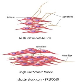
Termed unitary smooth muscle or visceral muscle, this type of smooth muscle is the most common observed in the human body, forming the walls of hollow organs.
The second type of muscle is the smooth muscle. Smooth muscle cells are found in the walls of hollow organs, including the stomach, intestines, urinary bladder and uterus, and in the walls of passageways, such as the. The calcium is the cause of protein to detach from the actin and myosin fastly binds with the opening of actin. Muscles of upper back 12 photos of the muscles of upper back map of upper back muscles, muscles of the upper back and chest, origin and insertion of upper back muscles, superficial muscles of the upper back, tight muscles of the upper back and neck, human muscles, map of upper back muscles, muscles of the … Smooth muscle, muscle that shows no cross stripes under microscopic magnification. • smooth muscles respond to stretch only briefly, and then adapts to its new length • the new length however, retains its original _____ seconds or minutes after it has been elongated or shortened (e.g. In addition, the contractile state of smooth muscle is controlled by hormones, autocrine/paracrine agents, and other local chemical signals. Diaphragm is also a skeletal muscle. It is the main muscle of respiration. The smooth muscles perform the functions in the contrast of other types of muscles. Smooth muscles, cardiac muscles and skeletal muscles. This smooth muscle can be found surrounding the walls of the blood vessels, the bronchioles in the lungs, and the sphincter muscles used in the gi tract.the gi tract, which is tubular by design, also houses longitudinal muscles in addition to the smooth. These cells have fibers of actin and myosin which run through the cell and are supported by a framework of other proteins.
Smooth endoplasmic reticulum also releases calcium ions when a cell receives nerve impulse and cause muscle contraction. Smooth muscles have a much stronger ability to contract than skeletal. Muscle cells, commonly known as myocytes, are the cells that make up muscle tissue.there are 3 types of muscle cells in the human body; There are three types of muscle in the body: Vascular smooth muscle is innervated primarily by the sympathetic nervous system through adrenergic receptors (adrenoceptors).
File Smooth Muscle Contraction1 Png Wikimedia Commons from upload.wikimedia.org Smooth muscle anatomy smooth muscle tissue is also known as visceral muscle tissue. It is layered in a distinctive pattern of circular layers. All skeletal muscle fibres are not the same. The smooth muscle, on the other hand, is found in the wall of blood vessels and viscera (for example in the wall of digestive tract). Related posts of smooth muscle diagram labeled muscle anatomy interactive. Smooth muscle fibers are often found forming sheets of tissue and function in a coordinated fashion due to the presence of gap junctions between the cells. Cardiac and skeletal myocytes are sometimes referred to as muscle fibers due to their long and fibrous shape. This smooth muscle can be found surrounding the walls of the blood vessels, the bronchioles in the lungs, and the sphincter muscles used in the gi tract.the gi tract, which is tubular by design, also houses longitudinal muscles in addition to the smooth.
Smooth muscle tissue, unlike striated muscle, contracts slowly and automatically.
The smooth muscle, on the other hand, is found in the wall of blood vessels and viscera (for example in the wall of digestive tract). There are three types of muscle in the body: It is layered in a distinctive pattern of circular layers. The second type of muscle is the smooth muscle. They are spindle shaped and have no striations. These muscles are often anchored by tendons. Smooth muscles, cardiac muscles and skeletal muscles. Smooth muscle cells lack the striated banding pattern found in cardiac and skeletal muscle, and they receive neural innervation from the autonomic nervous system. Smooth muscle tissue, unlike striated muscle, contracts slowly and automatically. It constitutes much of the musculature of In skeletal muscle, a single type of somatic nervous system traverses to muscle, where it stimulates organelle in the muscle cells in order to release calcium. In this video i am gonna to show you how to draw the diagrams of cardiac, straited, smooth muscle for class 1st to 10th. It is the pen diagram of skeletal, smooth and cardiac muscle for class 10, 11 and 12.
Muscles of upper back 12 photos of the muscles of upper back map of upper back muscles, muscles of the upper back and chest, origin and insertion of upper back muscles, superficial muscles of the upper back, tight muscles of the upper back and neck, human muscles, map of upper back muscles, muscles of the … It constitutes much of the musculature of It is layered in a distinctive pattern of circular layers. Smooth muscle anatomy smooth muscle tissue is also known as visceral muscle tissue. These are called contact junctions, and they function in much the same way as the skeletal.
Muscle The Histology Guide from www.histology.leeds.ac.uk Termed unitary smooth muscle or visceral muscle, this type of smooth muscle is the most common observed in the human body, forming the walls of hollow organs. These muscles are often anchored by tendons. Vascular smooth muscle is innervated primarily by the sympathetic nervous system through adrenergic receptors (adrenoceptors). Smooth muscle is composed of sheets or strands of smooth muscle cells. Cardiac and skeletal myocytes are sometimes referred to as muscle fibers due to their long and fibrous shape. It is also found in the smooth muscles cells of the body along with skeletal muscle cell but here it is more loosely organised compared to the smooth er of the skeletal muscle cells. Related posts of smooth muscle diagram muscles of upper back. The calcium is the cause of protein to detach from the actin and myosin fastly binds with the opening of actin.
Muscles of upper back 12 photos of the muscles of upper back map of upper back muscles, muscles of the upper back and chest, origin and insertion of upper back muscles, superficial muscles of the upper back, tight muscles of the upper back and neck, human muscles, map of upper back muscles, muscles of the …
The cardiac muscle is only found in the heart wall. In this video i have shown the simplest way of drawing muscle drawing. Smooth muscle makes up the walls of hollow organs, respiratory passageways, and blood vessels. In skeletal muscle, a single type of somatic nervous system traverses to muscle, where it stimulates organelle in the muscle cells in order to release calcium. Learn vocabulary, terms, and more with flashcards, games, and other study tools. Smooth muscle anatomy smooth muscle tissue is also known as visceral muscle tissue. Smooth muscle cells are found in the walls of hollow organs, including the stomach, intestines, urinary bladder and uterus, and in the walls of passageways, such as the. Vascular smooth muscle is innervated primarily by the sympathetic nervous system through adrenergic receptors (adrenoceptors). Muscle anatomy interactive 12 photos of the muscle anatomy interactive human muscle anatomy interactive, interactive muscle anatomy games, muscle anatomy interactive, muscle anatomy interactive quiz, shoulder muscle anatomy interactive, human muscles, human muscle anatomy interactive, interactive muscle anatomy. It is layered in a distinctive pattern of circular layers. Smooth muscles, cardiac muscles and skeletal muscles. Termed unitary smooth muscle or visceral muscle, this type of smooth muscle is the most common observed in the human body, forming the walls of hollow organs. Smooth muscles exhibits a phenomenon called _____ in which: